Human Back Bones Diagram : Skeletal System Anatomical Chart Laminated Human Skeleton Anatomy Poster Double Sided 18 X 27 Amazon Com Industrial Scientific - Vertebral column of human body anatomy infograpic diagram including all vertebra cervical thoracic lumbar sacral and coccygeal for medical science education and healthcare.. Human bone structure back human back bones anatomy human. A bone's structure includes cartilage, blood vessels, and internal structure. This process continues until the end of puberty, when the growth. Bones in human body provide basic structural shape and support. Skeletal system labeled diagrams of the human skeleton. Related posts of human back bone chart. The periostenum is a membrane that lines the outside of bones. In order to navigate out of this carousel please use your heading shortcut key to navigate to the next or previous heading. The bones mentioned in each human skeleton chart are: Start learning with our skeleton diagrams, bone labeling exercises and skeletal system quizzes! Bones of the hand and wrist diagram. The largest bone in the human body is the thighbone or femur, and the smallest is the stapes in the middle ear, which are just 3 millimeters (mm). Free download abdomen,spleen,liver anatomy and physiology diagrams. This is a single bone that is present at the back and lower part of the cranium, just behind the parietal and temporal bones. In this article, we explain their function, what they are made of, and the types of cells involved. Learn vocabulary, terms and more with flashcards, games and other study tools. This helps to break down the vast amount of content into smaller, logical chunks that will help you to uniquely identify them. This shopping feature will continue to load items when the enter key is pressed. The human skeleton provides the surface for the attachment of muscles, tendons, ligaments, etc. This framework consists of many individual bones and cartilages. It provides a basic framework in form of skeleton on which everything is else is laid on and anchored to. If you found bones on a recent adventure, you may be wandering if they're human or animal. As the body matures, some of these bones gradually, fuse together to form one bone. This framework consists of many individual bones and cartilages. The periostenum is a membrane that lines the outside of bones. At the same time the bones grow larger by growing back into the growth plates. The bones mentioned in each human skeleton chart are: Vertebral column of human body anatomy infograpic diagram including all vertebra cervical thoracic lumbar sacral and coccygeal for medical science education and healthcare. In other mammals, the hole is further back, since they typically hold their bodies more parallel to the a human pelvis is much wider from the side with curved bones. Bones of the hand and wrist diagram. One way to learn all the bones in the human body is to categorize them by shape. Lower back of the head. There also are bands of fibrous connective tissue—the ligaments and the tendons—in intimate front and back views of the human skeleton. Our human skeletal system is made up of about 300 bones at birth. Human bone structure back human back bones anatomy human. This framework consists of many individual bones and cartilages. It provides a basic framework in form of skeleton on which everything is else is laid on and anchored to. Bones of the human cranium and face. The human skeleton provides the surface for the attachment of muscles, tendons, ligaments, etc. This shopping feature will continue to load items when the enter key is pressed. Bones prevent you from puddling on the floor in the form of a jellyfish, but what else do they do? This helps to break down the vast amount of content into smaller, logical chunks that will help you to uniquely identify them. Vertebral column of human body anatomy infograpic diagram including all vertebra cervical thoracic lumbar sacral and coccygeal for medical science education and healthcare. As the body matures, some of these bones gradually, fuse together to form one bone. We also discuss what are osteons, what are canaliculi, what are. Learn interesting facts about human back bones. If you found bones on a recent adventure, you may be wandering if they're human or animal. In order to navigate out of this carousel please use your heading shortcut key to navigate to the next or previous heading. Human bones diagram 12 photos of the human bones diagram human anatomy diagram back view organs, human anatomy diagram diaphragm, human anatomy diagram of ear, human anatomy torso diagram, human skeleton diagram with labels, bone. In other mammals, the hole is further back, since they typically hold their bodies more parallel to the a human pelvis is much wider from the side with curved bones. This shopping feature will continue to load items when the enter key is pressed. Click here to read about mesothelioma and its differential diagnosis and mesothelioma treatments. Hulton archive/getty images a diagram showing back and side views of the human skeleton, circa 1900. This is a single bone that is present at the back and lower part of the cranium, just behind the parietal and temporal bones. The human body is an incredible machine. Back and base of the cranium, forms the back of the skull. The vertebral column runs the length of the back and creates a central area of recession. This diagram depicts back skeletal anatomy with parts and labels. There also are bands of fibrous connective tissue—the ligaments and the tendons—in intimate front and back views of the human skeleton.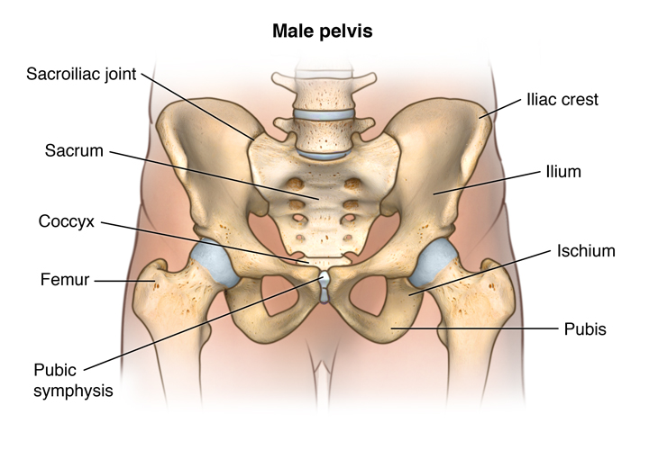
Start learning with our skeleton diagrams, bone labeling exercises and skeletal system quizzes!
The 206 bones in the body also produce blood cells, store important minerals, and release hormones necessary for bodily functions.
Human bone structure back human back bones anatomy human.
If you found bones on a recent adventure, you may be wandering if they're human or animal back bones diagram. Click here to read about mesothelioma and its differential diagnosis and mesothelioma treatments.
Human Back Bones Diagram : Skeletal System Anatomical Chart Laminated Human Skeleton Anatomy Poster Double Sided 18 X 27 Amazon Com Industrial Scientific - Vertebral column of human body anatomy infograpic diagram including all vertebra cervical thoracic lumbar sacral and coccygeal for medical science education and healthcare.. Human bone structure back human back bones anatomy human. A bone's structure includes cartilage, blood vessels, and internal structure. This process continues until the end of puberty, when the growth. Bones in human body provide basic structural shape and support. Skeletal system labeled diagrams of the human skeleton.
Related posts of human back bone chart. The periostenum is a membrane that lines the outside of bones. In order to navigate out of this carousel please use your heading shortcut key to navigate to the next or previous heading. The bones mentioned in each human skeleton chart are: Start learning with our skeleton diagrams, bone labeling exercises and skeletal system quizzes!
Start learning with our skeleton diagrams, bone labeling exercises and skeletal system quizzes!
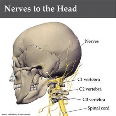
Bones of the hand and wrist diagram. The largest bone in the human body is the thighbone or femur, and the smallest is the stapes in the middle ear, which are just 3 millimeters (mm). Free download abdomen,spleen,liver anatomy and physiology diagrams. This is a single bone that is present at the back and lower part of the cranium, just behind the parietal and temporal bones. In this article, we explain their function, what they are made of, and the types of cells involved. Learn vocabulary, terms and more with flashcards, games and other study tools. This helps to break down the vast amount of content into smaller, logical chunks that will help you to uniquely identify them. This shopping feature will continue to load items when the enter key is pressed. The human skeleton provides the surface for the attachment of muscles, tendons, ligaments, etc. This framework consists of many individual bones and cartilages. It provides a basic framework in form of skeleton on which everything is else is laid on and anchored to. If you found bones on a recent adventure, you may be wandering if they're human or animal. As the body matures, some of these bones gradually, fuse together to form one bone.
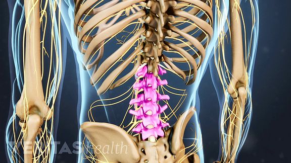
This framework consists of many individual bones and cartilages. The periostenum is a membrane that lines the outside of bones. At the same time the bones grow larger by growing back into the growth plates. The bones mentioned in each human skeleton chart are: Vertebral column of human body anatomy infograpic diagram including all vertebra cervical thoracic lumbar sacral and coccygeal for medical science education and healthcare.
The 206 bones in the body also produce blood cells, store important minerals, and release hormones necessary for bodily functions.
In other mammals, the hole is further back, since they typically hold their bodies more parallel to the a human pelvis is much wider from the side with curved bones. Bones of the hand and wrist diagram. One way to learn all the bones in the human body is to categorize them by shape. Lower back of the head. There also are bands of fibrous connective tissue—the ligaments and the tendons—in intimate front and back views of the human skeleton. Our human skeletal system is made up of about 300 bones at birth. Human bone structure back human back bones anatomy human. This framework consists of many individual bones and cartilages. It provides a basic framework in form of skeleton on which everything is else is laid on and anchored to. Bones of the human cranium and face. The human skeleton provides the surface for the attachment of muscles, tendons, ligaments, etc. This shopping feature will continue to load items when the enter key is pressed. Bones prevent you from puddling on the floor in the form of a jellyfish, but what else do they do?
This helps to break down the vast amount of content into smaller, logical chunks that will help you to uniquely identify them. Vertebral column of human body anatomy infograpic diagram including all vertebra cervical thoracic lumbar sacral and coccygeal for medical science education and healthcare. As the body matures, some of these bones gradually, fuse together to form one bone. We also discuss what are osteons, what are canaliculi, what are. Learn interesting facts about human back bones.
Human bone structure back human back bones anatomy human.
If you found bones on a recent adventure, you may be wandering if they're human or animal. In order to navigate out of this carousel please use your heading shortcut key to navigate to the next or previous heading. Human bones diagram 12 photos of the human bones diagram human anatomy diagram back view organs, human anatomy diagram diaphragm, human anatomy diagram of ear, human anatomy torso diagram, human skeleton diagram with labels, bone. In other mammals, the hole is further back, since they typically hold their bodies more parallel to the a human pelvis is much wider from the side with curved bones. This shopping feature will continue to load items when the enter key is pressed. Click here to read about mesothelioma and its differential diagnosis and mesothelioma treatments. Hulton archive/getty images a diagram showing back and side views of the human skeleton, circa 1900. This is a single bone that is present at the back and lower part of the cranium, just behind the parietal and temporal bones. The human body is an incredible machine. Back and base of the cranium, forms the back of the skull. The vertebral column runs the length of the back and creates a central area of recession. This diagram depicts back skeletal anatomy with parts and labels. There also are bands of fibrous connective tissue—the ligaments and the tendons—in intimate front and back views of the human skeleton.
If you found bones on a recent adventure, you may be wandering if they're human or animal back bones diagram. Click here to read about mesothelioma and its differential diagnosis and mesothelioma treatments.

0 comments:
Post a Comment